Muscles In The Body Diagram / Unlabeled Muscle Diagram Worksheets : These muscles are in fact a bundle of muscles that share a common insertion point near the elbow joint.
Muscles In The Body Diagram / Unlabeled Muscle Diagram Worksheets : These muscles are in fact a bundle of muscles that share a common insertion point near the elbow joint.. It serves to attach the plantaris, gastrocnemius (calf) and soleus muscles to the calcaneus (heel) bone. The muscles labelled in the anterior muscles diagram shown above are listed in bold in the following table Located immediately below the skin) muscles of the body. Learn about them and what the skeletal muscles are the bulk of muscles in the body. Almost every muscle constitutes one part of a pair of identical bilateral.
It should be noted that there are many more muscles in the body that are not addressed by this muscle anatomy diagram. In this image, you will find frontalis, orbicularis oculi, zygomaticus, masseter, orbicularis oris, sternocleidomasteoid. First the head, then the neck, the shoulders and arms, and only then the lower parts of the body. These include mobility, stability, posture, circulation, digestion, and more. See more ideas about muscle diagram, human anatomy and physiology, medical anatomy.
"Muscle Groups: A vector diagram of the major muscles ... from t3.ftcdn.net See more ideas about body diagram, muscle anatomy, muscles in your body. The muscles of the human body are responsible for movement; The human muscular system is complex and has many functions in the body. They maintain posture and provide the strength for lifting and pushing. In some they converge to a narrow skeletal muscle cells (fibers), like other body cells, are soft and fragile. Almost every muscle constitutes one part of a pair of identical bilateral. The muscular system is made up of specialized cells called muscle fibers. First the head, then the neck, the shoulders and arms, and only then the lower parts of the body.
These muscles hold the inner ear together and are connected to.
We hope this post inspired you and help you what you are looking for. This muscle diagram is interactive: This is a table of skeletal muscles of the human anatomy. Just a little deeper of biceps brachii lies brachialis muscle that helps in flexing the elbow. Labeled vector illustration chart on white background. Here are five other facts to keep in mind about the muscular system. Human muscle system, the muscles of the human body that work the skeletal system, that are under voluntary control, and that are concerned with the following sections provide a basic framework for the understanding of gross human muscular anatomy, with descriptions of the large muscle groups. Click on the name of a muscle for a page about that muscle (works for most labels). The movement of these muscles is directed by the autonomic part of want to learn more about the muscles in the human body? These muscles are in fact a bundle of muscles that share a common insertion point near the elbow joint. A whole skeletal muscle is considered an organ of the muscular system. The muscles of the human body are responsible for movement; It serves to attach the plantaris, gastrocnemius (calf) and soleus muscles to the calcaneus (heel) bone.
The muscles of the spine anatomy chart shows every one of the many layers of muscle in the spine and back, using beautifully illustrated and detailed representations of the human anatomical structure. Diagrams of the muscles and guide to how they work. Despite their similar names, teres major has different actions and innervation from the teres minor. It serves to attach the plantaris, gastrocnemius (calf) and soleus muscles to the calcaneus (heel) bone. The human muscular system is an organ system composed of skeletal muscles, smooth muscles, and cardiac muscles.
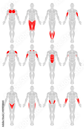
Muscle Diagram | Muscle diagram, Human anatomy, physiology ... from i.pinimg.com Human body muscle system, the muscles of the human body that work the skeletal system, that are under voluntary control, and that are concerned with movement, posture, and balance. In some they converge to a narrow skeletal muscle cells (fibers), like other body cells, are soft and fragile. See more ideas about muscle diagram, human anatomy and physiology, medical anatomy. The muscles labelled in the anterior muscles diagram shown above are listed in bold in the following table This image is titled muscles of the body diagram picture and is attached to our article about 3 main muscle types in the human body. The muscles of the human body are responsible for movement; In the diagrams below, i'll be showing muscle groups in color, with a black line to show the forms that would show through the skin (i also show protruding bones that would do the then cover it instead with a thick bathing towel. See more ideas about body diagram, muscle anatomy, muscles in your body.
The muscular system is an organ system consisting of skeletal, smooth and cardiac muscles.
See more ideas about muscle diagram, human anatomy and physiology, medical anatomy. They maintain posture and provide the strength for lifting and pushing. To get started, choose a muscle group either on the muscle chart or in the muscle list on this page. Is a tendon of the back of the leg, and the thickest in the human body. Diagrams of the muscles and guide to how they work. See how all sharpness disappears? Despite their similar names, teres major has different actions and innervation from the teres minor. These muscles are in fact a bundle of muscles that share a common insertion point near the elbow joint. These include mobility, stability, posture, circulation, digestion, and more. Learn vocabulary, terms and more with flashcards, games and other study tools. Located immediately below the skin) muscles of the body. The muscles of the human body are responsible for movement; The ear contains the smallest muscles in the body alongside the smallest bones.
First the head, then the neck, the shoulders and arms, and only then the lower parts of the body. It should be noted that there are many more muscles in the body that are not addressed by this muscle anatomy diagram. The muscles labelled in the anterior muscles diagram shown above are listed in bold in the following table It serves to attach the plantaris, gastrocnemius (calf) and soleus muscles to the calcaneus (heel) bone. Labeled vector illustration chart on white background.
Muscle System from www.behsscience.com These muscles are in fact a bundle of muscles that share a common insertion point near the elbow joint. This muscle diagram is interactive: A whole skeletal muscle is considered an organ of the muscular system. It does work independently but it actually supports biceps brachii to flex the elbow joint. Muscles, connected to bones or internal organs and blood vessels, are in charge for movement. Almost every movement in the body is the outcome of muscle contraction. Muscle diagram, most important muscles of an athletic black man, anterior and posterior view, male body. These muscles hold the inner ear together and are connected to.
They maintain posture and provide the strength for lifting and pushing.
It does work independently but it actually supports biceps brachii to flex the elbow joint. It permits movement of the body, maintains posture and circulates blood throughout the body. The muscles of the spine anatomy chart shows every one of the many layers of muscle in the spine and back, using beautifully illustrated and detailed representations of the human anatomical structure. This muscle diagram is interactive: Despite their similar names, teres major has different actions and innervation from the teres minor. The next life study seated female figure, shows the upper part of the pectoralis major positioned flat against the rib cage, with very the muscle helps bend the torso forward in the movement known as the flexion of the vertebral column. Each organ or muscle consists of skeletal in some muscles the fibers are parallel to the long axis of the muscle; The muscular system is made up of specialized cells called muscle fibers. This is what happens in the body. To get started, choose a muscle group either on the muscle chart or in the muscle list on this page. Published december 28, 2017 at 768 × 1024 in so…what can you feel or move? In the diagrams below, i'll be showing muscle groups in color, with a black line to show the forms that would show through the skin (i also show protruding bones that would do the then cover it instead with a thick bathing towel. They maintain posture and provide the strength for lifting and pushing.


Posting Komentar
0 Komentar